Aortic valve

Anatomy

The normal aortic valve consists of three equally sized valve cusps: the right coronary cusp (R), the left coronary cusp (L) and the non-coronary cusp (N). With 2D echocardiography, the valve can be assessed in the PLAX, where the RCC and the NCC are visible. In a normal tricuspid aortic valve, the closure line is (between RCC and NCC) usually in the centre of the aortic valve ring. However, in the PSAXao the three cusps in systole almost form a circle and during diastole the closure lines resemble an inverted Mercedes-Benz logo. Assessing the number of cusps is best done during systole, because the valve is open. When it’s closed, a so called raphe can be mistaken for a closing line. Today beautiful images of the aortic valve can be arrived with 3D echocardiography.

Aovalv

3D-image of a tricuspid aortic valve (TEE)

2D-image of a tricuspid aortic valve (TTE).

Regurgutation

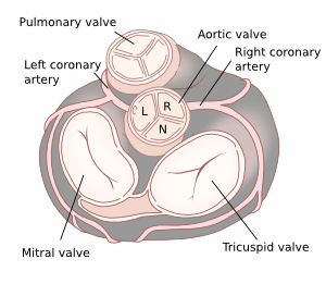
Quantification of aortic valve regurgitation

AR severity classes(a) Mild Moderate Moderate Severe
  grade 1 or 1+ Grade 2 or 2+ Grade 3 or 3+ Grade 4 or 4+
Qualitative parameters
Aortic valve morphology Normal/abnormal Normal/abnormal Abnormal/prolaps/moderate coaptation defect Abnormal/flail/lage coaptation defect
Colour flow AR jet width(b) Small in central jet Intermediate Large in central jet, variable in eccentric jets Large in central jet, variable in eccentric jets
Colour flow convergence None or very small Intermediate Intermediate Large
CW signal of AR jet Incomplete/faint Dense Dense Dense
Diastolic flow reversal in descending aorta Brief, proto-diastolic flow reversal Intermediate Holodiastolic flow reversal Holodiastolic flow reversal
      end-diastolic velocity 10 to <20 cm/s end-diastolic velocity ≥20 cm/s
Diastolic flow reversal in abdominal aorta Absent Absent Present Present
Semi-quantitative parameters
Vena contracta (mm) <3 3-6 3-6 >6
Jet width/LVOT diameter (%) <25 25-45 46-64 >65
Jet CSA / LVOT CSA (%) <5 5-20 21-59 ≥60
Pressure half time (m/s)(c,d) >500 500-200 500-200 <200
Quantitative parameters
EROA (mm²) <10 10-19 20-29 ≥30
R Vol (mL) <30 30-44 45-59 ≥60
RF (%) <30 30-39 40-49 ≥50
Structural parameters
LV size(e) Usually normal Normal or dilated usually dilated usually dilated

AR, aortic regurgitation; CSA, cross-sectionnal area; CW, continuous wave; LA, left atrium; EROA, effective regurgitant orifice area; LV, left ventricle; RF, regurgitant fraction; R Vol, regurgitant volume; VC, vena contracta.
(a)Grading of severity of AR classifies regurgitation as mild, moderate or severe, and sub-classifies the moderate regurgitation group into two subclasses: ‘mild-to-moderate’ (EROA of 10–19 mm or a R Vol of 20–44 mL) and ‘moderate-to-severe’(EROA of 20–29mm or a R Vol of 45–59mL).
(b)At a Nyquist limit of 50–60 cm/s.
(c)These parameters are influenced by LV and aortic compliance. Hence, low transvalvular end-diastolic aorta to LV pressure gradient due to concomitant moderate/severe LV diastolic dysfunction may lead to false positive results. The high dependency of aortic flow reversal on aortic compliance considerably limits the utility of this parameter in the elderly population. These parameters are also influenced by chronotropy.
(d)Pressure half-time is shortened with increasing LV diastolic pressure, vasodilator therapy, and in patients with a dilated compliant aorta or lengthened in chronic AR.
(e)Unless for other reasons, the LV size is usually normal in patients with mild AR. In acute severe AR, the LV size is often normal. Accepted cut-off values for non-significant LV enlargement: LV end-diastolic volume <82 mL/m2, LV end-systolic diameter <40 mm2, LV end-systolic volume <30 mL/m2.

Causes of aortic valve regurgitation

Causes
Congenital Bicuspid aortic valve.
Damage to the aortic valve Rheumatic fever.
  Degeneracy.
  Endocarditis.
  Syphilis.
  Trauma.
  Metabolic diseases such as mucopolysaccharidosis.
Aortic dilation Connective tissue disease, such as Marfan's syndrome, Ehlers-Danlos' syndrome.
  Aneurysm by inflammation, such as syphilis, vasculitis.
  Aneurysm in aortic atherosclerosis.
  Aortic dissection.

Stenosis

Quantification of aortic valve stenosis

Parameter Mild Moderate Severe
PG max (mmHg) 15-40 40-70 70-100
PG mean (mmHg) <20 20-40 >40
Acceleration time (ms)     >100
Vmax (m/s) <3 3-4 >4
AVA (cm²) >1.5 1.5-1.0 <1.0
AVA BSAindex (cm²/m²) >0.85 0.85-0.60 <0.60
Dimensionless index > 0.50 0.25-0.50 <0.25

Causes of aortic valve stenosis

Supravalvular Valvular Subvalvular
A pleat or membrane of the aorta Congenital form; often the aortic valve is bicuspid Congenital malformation: a membrane in LVOT
Narrowing of the aorta By rheumatic fever Hypertrophic obstructive cardiomyopathy, an autosomal dominant hereditary disorder
Williams Syndrome degenerative form Subvalvular aortic stenosis has also been described as a result of any of the lysosomal storage diseases


Bicuspid

Bicuspid aortic valve (BAV) is the most common congenital cardiac valve abnormality. In BAV two valve leaflets are fused from birth. BAV occurs in 1-2% of the population and twice as often in men as in women. BAV can be hereditary in which both familial clustering and isolated valve defects are documented. The incidence of bicuspid aortic valve may reach up to 10% in certain families. A BAV is often associated with other congenital heart defects such as coarctation of the aorta. Usually there is a fusion between the LCC and RCC (70%). Initially, the bicuspid aortic valve will not cause problems. Later in life, the valve has an increase risk of calcified deformation which can lead to stenosis and/or regurgitation through malcoaptation of the affected cusps. In BAV cusps are of unequal size and usually show a raphe at the largest cusp. A raphe is a fibrotic ledge that connects the edges of congenital fused cusps and does not consist of valve tissue.

bav

Bicuspid type 2

Bicuspid type 2

The information above comes from Echocardiografie.nl. Last changed on: 7 September 2023.