In echocardiography there are a number of standard positions in which images of the heart can be made. These so-called acoustic windows are only recordable between/below and / or above the ribs. Every sonographer must reckon the fact that not every patient’s heart is actually in the same position to be imaged. Sometimes it is necessary to scan an intercostal space higher or lower, or more medial or lateral. Also, not every patient equally absorb the ultrasound waves for example fat (obesity)) or air (COPD) will require a lot of creativity and improvisational skills of the sonographer. It is advisable to operate according to the same protocol so all the information is delivered to the assessor in order and there is less chance of forgetting recordings and / or measurements.
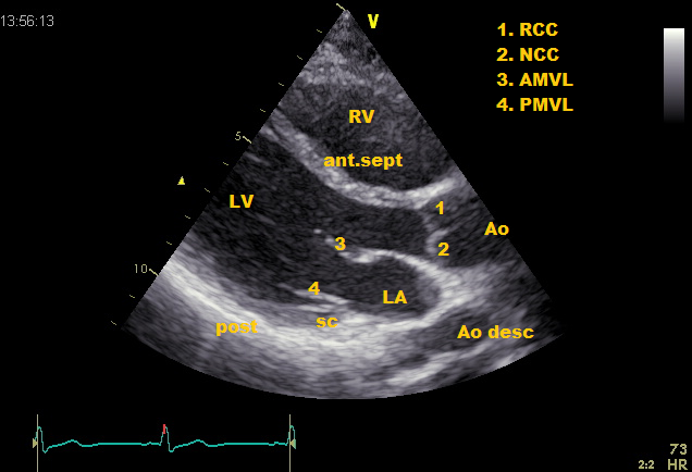
Parasternal long axis (PLAX)
Place the transducer on the 3rd intercostal space left parasternal. The transducer index mark is directed to the right shoulder of the patient. The right (RV) and left ventricle (LV), as well as the aortic root (Ao), and the left atrium (LA) are displayed. Other structures which can be seen in this recording are: Pericardium, anterior wall of the right ventricle, right ventricular outflow tract, ascending aorta, sinuses of Valsalva, right coronary cusp and the non / left coronary cusp of the aortic valve, the anterior mitral valve leaflet and posterior mitral valve leaflet, left ventricle, left ventricular outflow tract. Membranous septum, muscular septum, coronary sinus, descending aorta (round shape under LA). In some cases, pericardial effusion, or pleural effusion.
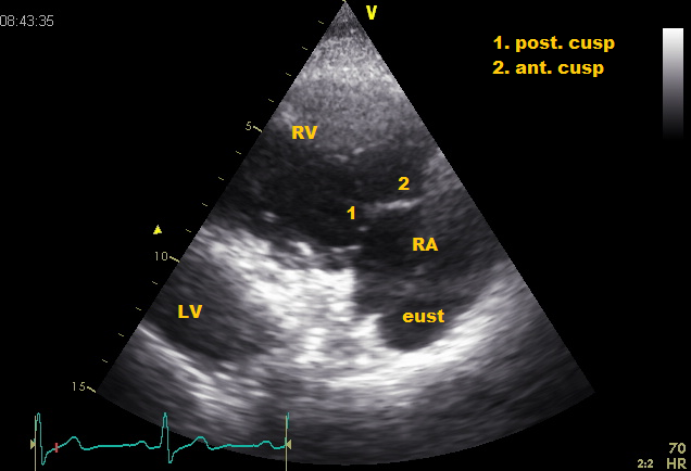
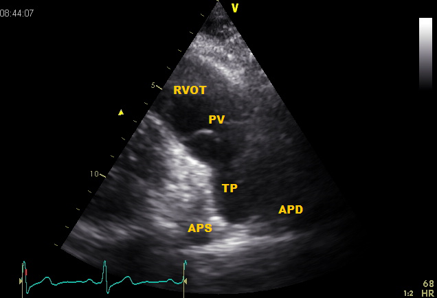
Parasternal long axis tilted
Tricuspid level
From PLAX tilt inferiorly. This will make the left-hand half of the heart is turned away, and the right ventricle (RV) and the right atrium (RA) at the edges. Also, the tricuspid valve is here clearly visible especially the posterior and the anterior cusp.
Pulmonary level
From PLAX tilt to cranial. This is a great image for the Pulmonary valve (PV), pulmonary artery, including the pulmonary trunk (TP) which splits into the right (APD) and left pulmonary artery (APS).
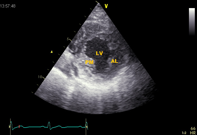
Parasternal short axis (PSAX)
Papillary level
turn the transducer 90° clockwise from the PLAX view. The left ventricle (LV) at the level of the papillary muscles, and the right ventricle (RV) are shown.
Mitral level
Lift the transducer a little to cranial from the same position. A cross section of the left ventricle (LV) at the level of the mitral valve and the right ventricle (RV) are shown. Other structures which can be seen in this recording are: Pericardium, anterior wall of the right ventricle, right ventricle, interventricular septum, left ventricular outflow tract, anterior mitral valve leaflet, posterior mitral valve leaflet, lateral mitral commissure and the medial mitral commissure, descending aorta.
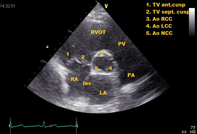
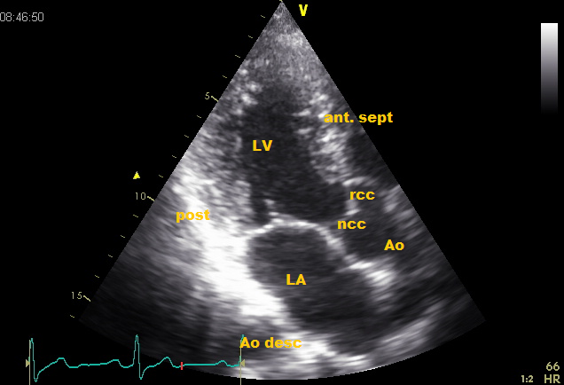
Aortic valve level
Lift the transducer a little more cranial from the same position . The aortic valve (AV), pulmonary valve (PV), left atrium (LA), right atrium (RA), tricuspid valve (TV) and the right ventricular outflow tract (RVOT) are displayed. Other structures that can be seen in this recording are: Pericardium, anterior wall of the right ventricle, pulmonary valve annulus, pulmonary trunk, right and left pulmonary artery, sinuses of Valsalva, left-, right-, and noncoronary-cusp of the aortic valve, inferior vena cava and the atrial septum.
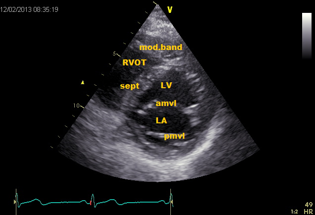
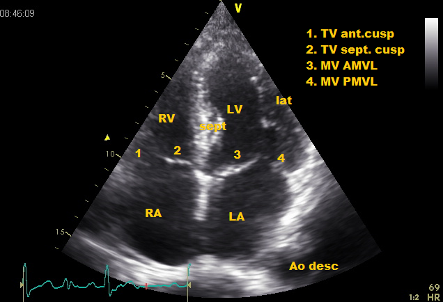
Apical four chamber view (AP4CH)
Place the transducer on the left 5th intercostal space, midclavicular. The transducer index mark is directed to the left side of the patient. The left (LV) and the right ventricle (RV), as well as the left (LA) and the right atrium (RA) can be displayed. Other structures that can be seen in this image are: left and right upper pulmonary veins, anterior tricuspid valve leaflet, septal tricuspid valve leaflet, moderator band, atrial septum, interventricular septum, descending aorta, pericardium.
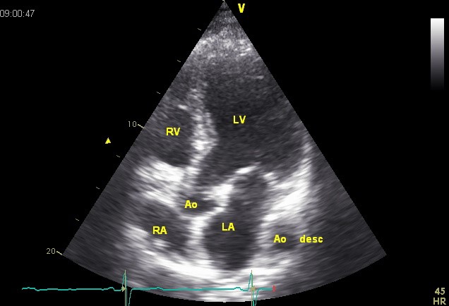
Apical five chamber view (AP5CH)
Create a four chamber view and then tilt the transducer slightly lower. The aortic valve (Ao) is shown in the middle between the chambers. Tilting of the transducer to the other direction, the coronary sinus can be displayed. Other structures which can be seen in this recording are: ascending aorta, aortic root, descending aorta, sinuses of Valsalva, right-, and noncoronary-cusp of the aortic valve and the left ventricular outflow tract.
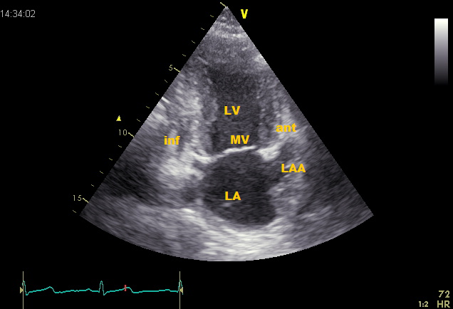
Apical two chamber view (AP2CH)
Turn the transducer approximately 60° counter- clockwise from the AP4CH position. The left ventricle (LV) and the left atrium (LA) can be displayed. Other structures which can be seen in this recording are: Pulmonary veins and the left atrium appendix (LAA).
Apical three chamber view (AP3CH)
Turn the transducer further, about 60° counter-clockwise from the AP2CH position and tilt slightly to the side. The left ventricle (LV), left atrium (LA) and the aortic root (Ao) are displayed.
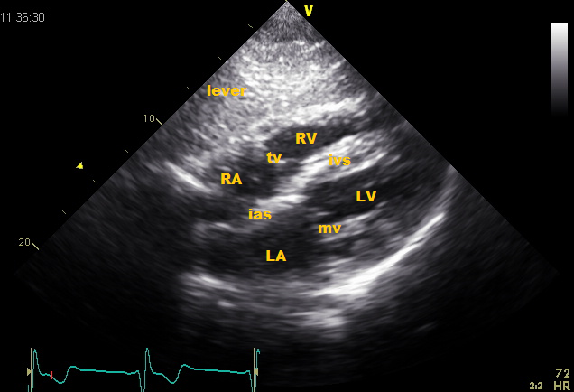
Subcostal view
Subcostal four chamber view (Subcostal 4CH)
Place the transducer on the subxyphoid region. The liver (L), left ventricle (LV), right ventricle (RV), left atrium (LA) and the right atrium (RA) are displayed. In this tilted AP4CH view the interventricular septum and atrial septum are almost perpendicular affected so this recording is very suitable to demonstrate a possible shunt (ASD/PFO or VSD).
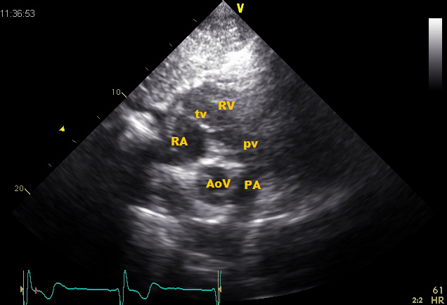
Subcostal SAX
Turn the transducer 90° clockwise relative to the subcostal 4CH view. The aortic valve (AOV), pulmonary valve (PV), right atrium (RA), tricuspid valve (TV) and the right ventricular outflow tract (RVOT) are shown. Other structures are pulmonary trunk, right and left pulmonary artery, sinuses of Valsalva, left-, right-, and noncoronary-cusp of the aortic valve, and atrial septum.
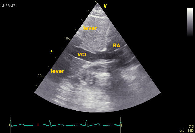
Subcostal IVC
Turn the transducer 45° counter clockwise relative to the subcostal 4CH view and tilt some. The transducer index mark facing the head of the patient. The inferior vena cava (IVC) is displayed. Turn the transducer slightly to the right again and the right and left ventricle (LV), and the right atrium (RA) and left atrium (LA) shown. This image shows a portion of the liver and is very suitable for the evaluation of right atrium, pericardial effusion and / or pleural fluid, and hepatic veins.
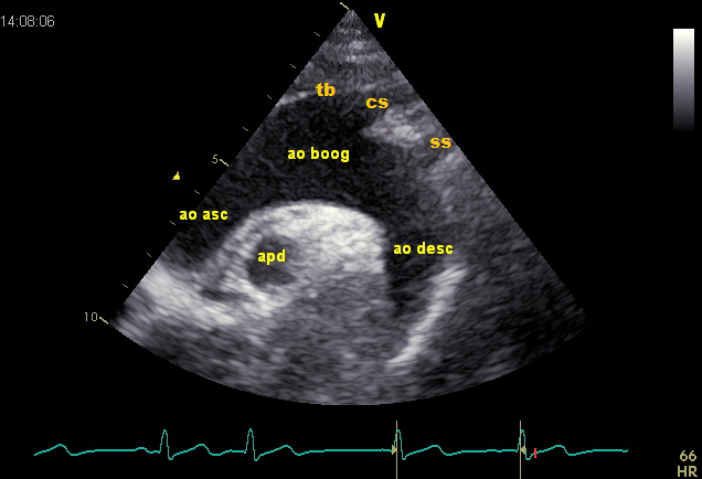
Suprasternal notch window
Place the transducer above the sternal jugular region. The transducer index mark facing the head of the patient and rotate approximately 45° to the right. This recording provides information about the ascending aorta (Ao asc), aortic arch (AO boog) with the right brachiocephalic (BC), the left common carotid (CC), subclavian artery (SS) and the descending aorta (Ao desc). Beneath the aortic arch a transverse cross-sectional view of the right pulmonary artery (APD) is seen, with the left atrium (LA) lying inferior.
The information above comes from Echocardiografie.nl. Last changed on: 7 September 2023.