This is a congenital heart defect in which blood flows between the atria of the heart. Normally, the atria are separated by the interatrial septum. If this septum is defective or absent, then oxygen-rich blood can flow directly from the left side of the heart to mix with the oxygen-poor blood in the right side of the heart, or vice versa. This can lead to lower-than-normal oxygen levels in the arterial blood that supplies the brain, organs, and tissues. However, an ASD may not produce noticeable signs or symptoms, especially if the defect is small. ASD can manifest at any age. An ASD is presented in 9% of all congenital heart diseases, but is the most common congenital heart disease which often reflects in adulthood (30%).
Types of ASD
| Type | Name | Location |
|---|---|---|
| ASD type I | Ostium primum defect | At the level of atrioventricular transition |
| ASD type II | Ostium secundum defect | In the middle of the atrial septum, in the fossa ovalis area |
| ASD type III | Sinus venosus defect | At the entrance of both the SVC and the IVC |
| Coronary sinus defect | Coronary sinus defect | Compound coronary sinus with LA |
Prevalence of ASD
| Type | Percentage |
|---|---|
| ASD II | 0.7 |
| ASD I | 0.15 |
| Sinus venosus defect | 0.1 |
| Coronary sinus defect | Rare |
Patent foramen ovale (PFO)
During development of the fetus, the interatrial septum develops to separate the left and right atria. However, a hole in the septum called the foramen ovale, allows blood from the right atrium to enter the left atrium during fetal development. This opening allows blood to bypass the nonfunctional fetal lungs while the fetus obtains its oxygen from the placenta. A layer of tissue called the septum primum acts as a valve over the foramen ovale during fetal development. After birth, the pressure in the right side of the heart drops as the lungs open and begin working, causing the foramen ovale to close entirely. In approximately 25% of adults, the foramen ovale does not entirely seal. In these cases, any elevation of the pressure in the pulmonary circulatory system can cause the foramen ovale to remain open. This is known as a patent foramen ovale (PFO), a type of atrial septal defect.
A PFO is involved with a number of other pathologies such as cerebrovascular accident (CVA), Transient Ischemic Attack (TIA), Caisson Disease (Decompression sickness in divers), Platypnoe-orthodeoxysyndrome and Migraine. There is a direct relationship between the size of the shunt and the risk of having a stroke.
A PFO can be demonstrated with the aid of contrast, by means of a saline solution. A PFO indicates a crossing from right to left of contrast within three cardiac cycles after injection.
Size of PFO
| Localization of defect | Indications for volume load | Indications for pulmonary hypertension |
|---|---|---|
| Where in IAS? | Indication of dilatation? | Indication of high pressures? |
| Color Doppler | Dimensions of RV | Systolic PA pressure arrived from TRmaxPG |
| Additional saline agitation | Dimensions of RA | Acceleration time in RVOT |
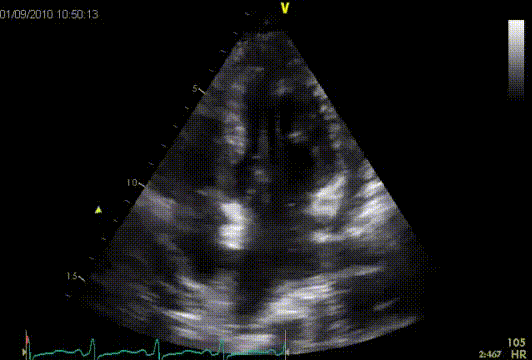
Large ASD II subcostal view
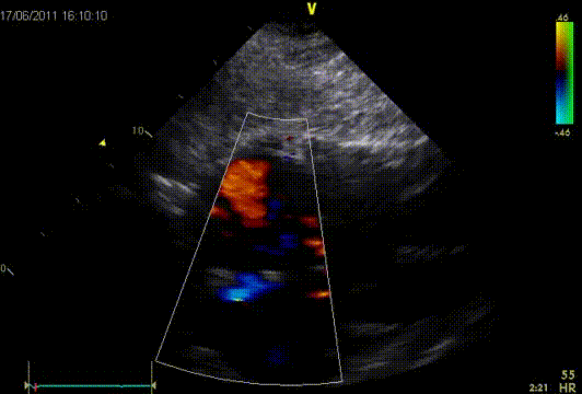
ASD II demonstrated with saline agitation
The information above comes from Echocardiografie.nl. Last changed on: 6 September 2023.