Persistent left superior vena cava

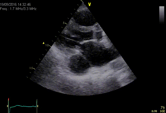
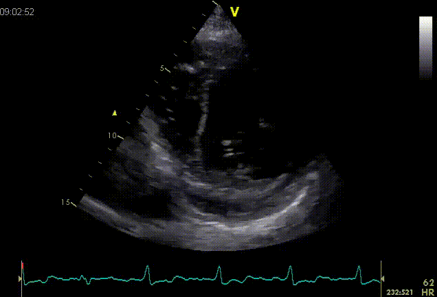
Persistent left superior vena cava (PLSVC) results from failure of obliteration of the left common cardinal vein, and it typically drains the left jugular and subclavian veins into the right atrium via the coronary sinus. It is the most common variant of thoracic venous drainage, and it is present in 0.5% of the general population. Its incidence increases to 3-10% in patients with congenital heart disease. In 80-90% of the cases a co-existent right superior vena cava is also present, although it can be smaller than usual. Besides coronary sinus, other drainage sites are also possible. In less than 10 percent the persistent PLSVC drains directly into the left atrium or in a pulmonary vein. This type of drainage is almost always associated with other congenital anomalies. It results in right to left shunt and has been associated with cyanosis, paradoxical embolism or brain abscess. A PLSVC is often an incidental finding during transthoracic echocardiography. It is diagnosed indirectly through recognition of a dilated coronary sinus in parasternal long axis view. The coronary sinus is a circular structure in the atrioventricular groove, located anterior to the pericardium. A dilated coronary sinus must be differentiated from descending aorta, pericardial effusion or pulmonary vein. From four-chamber view with posterior angulation coronary sinus can be viewed in the long axis passing behind the left atrium towards the right atrium.

VCSSP

The differential diagnosis of coronary sinus dilatation includes:

  • Persistence of left superior vena cava.
  • Any cause of elevated right atrial pressure.
  • Partial anomalous pulmonary venous drainage.
  • Coronary arterio-venous fistula.
  • Unroofed coronary sinus with shunt flow between left atrium and coronary sinus.

In case of a dilated coronary sinus contrast injection in the antecubital veins can help establish the diagnosis of a PLSVC. Contrast echocardiography from the left antecubital vein will classically show early coronary sinus opacification, before the right atrium and right ventricle. Imaging is usually done from the parasternal long axis view. In the rare cases of left atrial drainage contrast injection in the left arm will determine opacification of the left atrium. Right antecubital vein contrast injection is followed by a normal sequence of opacification starting from the right atrium and followed by the right ventricle, with no contrast in the coronary sinus.

VCSSP02

Echocardiographic criteria for PLSVC diagnosis are:

  • Dilated coronary sinus in the absence of elevated right side filling pressure.
  • Coronary sinus opacification before the right atrium when contrast (?bubble study?) is injected in the left antecubital vein.
  • Normal sequence of opacification after right arm antecubital vein injection.

The information above comes from Echocardiografie.nl. Last changed on: 6 September 2023.