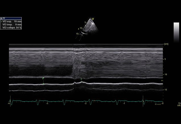
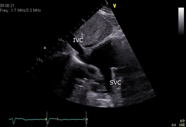
Inferior vena cava (IVC)
The inferior vena cava (IVC) is the body’s largest vein that provides the back-flow of blood to the heart. This vein contains deoxygenated blood returning from the tissues in the lower part of the body (systemic circulation). The inferior vena cava can be best appreciated from a subcostal view. The IVC can be compressed by surrounding tissues such as tumours obstructing the return of blood leading to congestion. This is called the IVC-Syndrome.
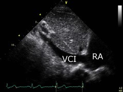
Inferior vena cava diameter
Normal | Borderline | Dilated |
---|---|---|
<15mm | 15-20mm | >20mm |
Respiratory collaps
With normal RA pressures the IVC collapses in 80% of cases, >40% (measured at the origin of the hepatic vein). 40% collapse corresponds to a pressure of 8-10mmHg in the IVC. At a collapse of 60-70%, the pressure is usually in the IVC 2-4mmHg. With a collapse <40% of the IVC, the pressures exceed >10mmHg. When the IVC is dilated and does not collapse, IVC the pressures can reach 20-25mmHg.

RA pressure
RA estimated pressure | ||
---|---|---|
Inferior Vena Cava size (mm) | IVC collaps (%) | Estimation RA pressure (mmHg) |
<21 | >50 | 3 (range 0–5) |
<21 | < 50 | 8 (range 5–10) |
>21 | >50 | 8 (range 5–10) |
>21 | < 50 | 15 (range 10- 20) |
Flow patterns
Because in most cases the the flow in the IVC is perpendicular to the ultrasound-beam, it can not be measured by Doppler reliably. In stead the flow pattern of the hepatic vein can be recorded. They mark the same signals, although the quality of the latter signal is much better because its flow is parallel to the sound beam.

Superior vena cava (SVC)
The superior vena cava contains the deoxygenated blood returning from the tissues in the upper part of the body. The superior vena cava connects into the roof of the right atrium. The superior vena cava can transthoracically not be portrayed properly. Some times it can be visualized supraclavicular, however this is not a default recording. The SVC (the lower part) is easier to visualize with TOE. The SVC can be compressed by tumours obstructing the return of blood leading to congestion, this is called the SVC-syndrome.

References
Res Cardiovasc Med. 2015 Aug 1;4(3):e28913 Eur Heart J Cardiovasc Imaging. 2015 Mar;16(3):233-70