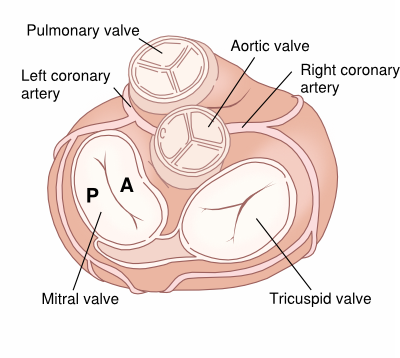
Anatomy
The mitral valve consists of two leaflets, the anterior (A) and the posterior valve leaflet (P), which together have a surface area of 4-6cm. At the inner edges of the leaflets chordae tendinae are attached, which ensure that the leaflets did not prolapse back into the atria during systole. The chordae attach to two large papillary muscles, that are part of the muscular tissue of the left ventricle.

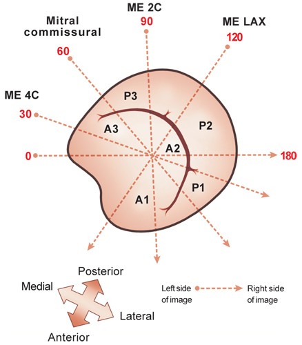
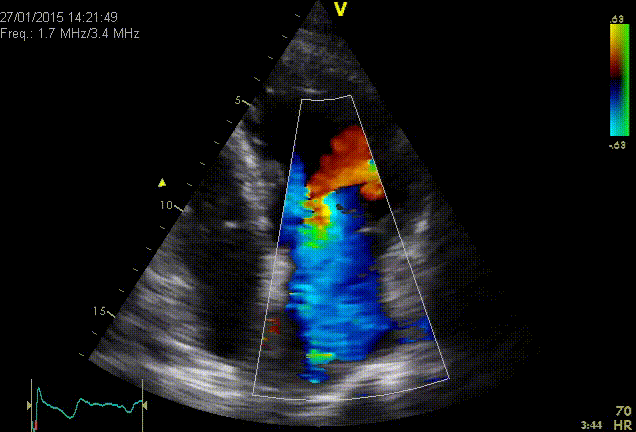
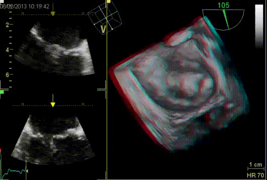
The mitral valve can be visualized in many views: PLAX, PSAXmv, AP4Ch, AP5Ch, AP2Ch, AP3Ch and subcostaal4Ch. A prolapse of the MV is best assessed on PLAX. However, mitral regurgitation should be assessed in all views. In case of major abnormalities of the mitral valve 3D TEE has additional value in assessing the anatomy and function.
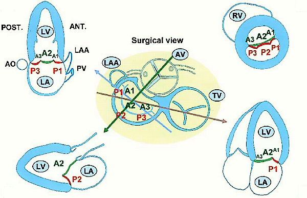
Scallop partition of mitral valve
Regurgitation
Quantification of mitral valve regurgitation
Grading severity mitral regurgitation | ||||
---|---|---|---|---|
MR severity classes 𝜁 | Mild | Moderate | Moderate | Severe |
Grade 1 or 1+ | Grade 2 or 2+ | Grade 3 or 3+ | Grade 4 or 4+ | |
Qualitative | ||||
Valve morphology | None or mild leaflet abnormality | Moderate leaflet abnormality | Moderate leaflet abnormality | Flail leaflet / large coaptation defect |
or minimal tenting | or moderate tenting | or moderate tenting | or severe tenting | |
Colour flow MR jet | Small, central | Intermediate | Intermediate | Very large central jet or eccentric jet adhering, |
swirling and reaching the posterior wall of the LA | ||||
Usually <4cm² | Usually 4-6cm² | Usually 6-8cm² | Usually >8cm² | |
<20% of LA | 20-30% of LA | 30-40% of LA | >50% of LA | |
Flow convergence zone 𝛼 | No or small faint | Dense | Dense | Large throughout systole, dense, holosystolic |
CW signal of MR jet | Parabolic | Partial or parabolic | Parabolic or triangular | Triangular |
Semi quantitative | ||||
Vena contracta width (mm) | <3 | 3 to <5 | 5 to <7 | ≥7 (>8 for biplane) 𝛽 |
Pulmonary vein flow | Systolic dominance | Variable | Systolic blunting | Systolic pulmonary vein flow reversal 𝛾 |
MV inflow | A wave dominant 𝛽 | Variable | E-wave dominant (E>1.2m/s) 𝜀 | E-wave dominant (E>1.2m/s) 𝜀 |
VTI mitral / VTI LVOT | <1 | Intermediate | >1.2 𝜀 | >1.4 𝜀 |
Quantative | ||||
EROA (mm²) | <20 | 20-29 | 30-39 | ≥40 |
R Vol (mL) | <30 | 30-44 | 45-59 | ≥60 |
RF (%) | <30 | 30-39 | 40-49 | ≥50 |
Structural parameters | ||||
LV and LA size | Usually normal | Normal or dilated | Usually dilated | Usually dilated |
PA pressure | Usually normal | Normal or elevated | Normal or elevated | Usually elevated |
CW, continuous wave; EROA, effective regurgitant orifice area; LA, left atrium; LV left ventricle; MR, mitral regurgitation; PA pulmonary artery; RF, regurgitant fraction; R Vol regurgitant volume; VC, vena contracta. 𝛼 At Nyquist limit of 50-60 cm/s. 𝛽 For average between AP4Ch an AP2Ch. 𝛾 Unless other reasons, of systolic blunting (atrial fibrillation, elevated LA pressure). 𝛿 Usually after 50 years of age. 𝜀 In the absence of other causes of elevated LA pressure and of mitral stenosis. 𝜁 Different thresholds are used in secondary mitral regurgitation where an EROA >20mm² and regurgitant volume > 30mL identify a subset of patients at increased risk of cardiac events. |
Echocardiographic criteria for the definition of severe mitral valve regurgitation: an integrative approuch
Parameter | Criteria | |
---|---|---|
Qualitative | ||
Valve morphology | Flail leaflet/ruptured pappilairy muscle/large coaptation defect | |
Colour flow regurgitant jet | Very large central jet or eccentric jet adhering, swirling and reaching the posterior wall of the LA | |
CW signal of regurgitant jet | Dense/triangular | |
Other | Large flow convergence zone * | |
Semiquantitative | ||
Vena contracta width (mm) | ≥7 (>8 for biplane) ** | |
Upstream vein flow | Systolic pulmonary vein flow reversal | |
Inflow | E-wave dominant ≥1.5 m/s *** | |
Other | TVI mitral/TVI aortic >1.4 | |
Quantative | Primary | Secundary **** |
EROA (mm²) | ≥40 | ≥20 |
Regugitant volume (mL/beat) | ≥60 | ≥30 |
Enlargement of cardiac chamber/vessels | LV, LA | |
* At Nyquist limit of 50-60 cm/s. ** For average between AP4Ch an AP2Ch. *** In the absence of other causes of elevated LA pressure and of mitral stenosis. **** Different thresholds are used in secondary mitral regurgitation where an EROA >20mm² and regurgitant volume > 30mL identify a subset of patients at increased risk of cardiac events. |
Severe MR because of prolaps
Causes of mitral valve regurgitation
Annulus dilation | Degeneracy | Acquired valve deviation | Secundary |
---|---|---|---|
Atriumfibrillation | Malcaptation in mitral valve stenosis | Billowing | Rhythm disorders (LBBB-->diastolic MR) |
LV dilation | Prolaps | SAM in HCM | |
LA dilation | Floppy valve | ||
Flail leaflet | |||
Chordal rupture | |||
Ruptured Papillary muscle |
Ruptured mitral chordae tendineae assessed with 3D TEE
Stenosis
Quantification of mitral stenosis
Parameter | Mild | Moderate | Severe |
---|---|---|---|
MVA (cm²) | > 1.5 | 1.5 - 1.0 | < 1.0 |
PGmean (mmHg) | < 5 | 5 - 10 | > 10 |
PHT (m/s) | < 90 | 90 - 150 | > 150 |
Causes of mitral stenosis
Acquired | Tumor | Congenital |
---|---|---|
Degeneracy | Myxoma | Parachute valve |
Rheumatic fever | Cor triatriatum sinistrum |
References
European Heart Journal - Cardiovascular Imaging (2022) 23, e171–e232 J Am Soc Echocardiogr. 2009 Jan;22(1):1-23; quiz 101-2 Eur J Echocardiogr. 2010 May;11(4):307-32 European Heart Journal (2017) 00, 1-53