Pericarditis

Pericarditis is an inflammation of the pericardium (heart sac) usually caused by a virus infection. The echocardiogram may show pericardial effusion of any size, pericardial thickening or a normal pericardium. A cardiac tamponade is a feared complication of pericarditis. This occurs when the pressure in the pericardium exceeds the pressure in the cardiac chambers, resulting in impaired cardiac filling and depends on the volume and rate of fluid accumulation.

Tamponade

Echocardiographic findings of pericarditis

Modality Findings
M-mode Rapid early diastolic and flat mid-diastolic movement of LV and aortic root.
Early diastolic notch in movement pattern of interventricular septum.
End-diastolic notch, followed by forward movement of septum.
2D images Thickened Pericardium (often difficult to detect, much more reliable MRI).
Septal motion to LV at inspiration (AP4CH).
Septal motion to RV at expiration (AP4CH).
Early opening of pulmonary valve.
Gedilateerde vena cava inferior met nauwelijks respiratoire variatie.

Inflow obstruction

With inspiration an increase of RV volume (shift in septal motion towards the LV in diastole and towards the RV in systole) and a decrease during expiration is seen. This is called interdependence. Due to inspiration the intrathoracic pressure becomes more negative and enhanced RV filling limits the LV diastolic filling. This corresponds with the physical finding of pulsus paradoxus.

Echocardiographic findings in inflow obstruction

Modality Findings
Doppler Increased respiratory variation in peak velocity of mitral valve (> 25%) and tricuspid valve (> 40%)
2-D images Dilated inferior vena cava without respiratory variation in diameter
Late diastolic to early systolic collapse of right and left atrium
Early diastolic collaps of right ventricle

Doppler measurements

The RV early diastolic filling velocity is augmented with inspiration, while the LV diastolic filling velocity is diminished. The E/A ratio of the mitral and tricuspid-inflow pattern will be >1. The deceleration time of the mitral E-wave will also be shortened (>180 msec.) In the pulmonary vein flow, the S-wave will be smaller than the D-wave.?This is different in a constrictive pericarditis. A respiratory variation in MV inflow (at at the beginning of inspiration increase of the E-wave >25%) and TV inflow (at the beginning of inspiration increase of the E-wave >40%) is seen. Also a respiratory variation in the pulmonary vein is seen, in which the flow at the beginning of expiration, the diastolic forward flow increases >10%. This respiratory variation can also be observed at atrial fibrillation and in patients with severe obstructive lung disease. In these cases the flow from the vena cava towards the RV will increase However in a constrictive pericarditis, the flow will not increase. After a pericardectomy an abnormal filling pattern is often observed. It is believed that this is due to incomplete pericardectomy. On the other hand the myocardium sometimes is involved in the disease process. The early diastolic velocity of the septal mitral valve annulus (é) is often reduced postoperatively, which indicates a relaxation disorder of the myocardium. This could be explained by increased myocardial fibrosis found at autopsies. Development of constrictive pericarditis after pericardectomy is associated with a worse prognosis.

PCinflowvariati1
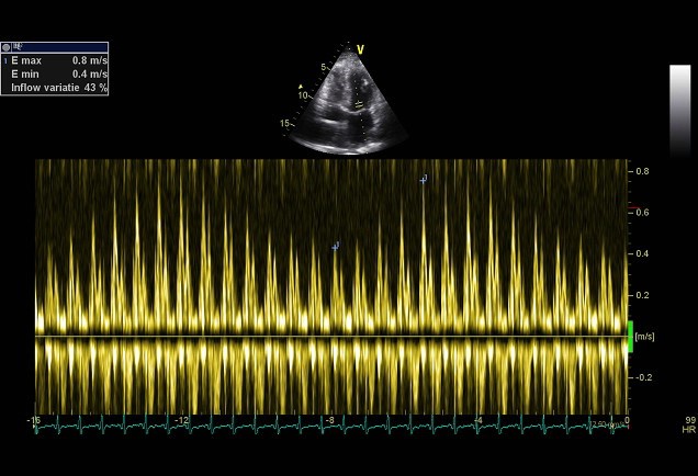
Respiratory flow variation >25%

Constrictive pericarditis


Variable Sensitivity, % Specificity, % Positive Predictive Value, % Negative Predictive Value, %
Individual variables
No. 1 ventricular septal shift 93 69 92 74
No. 2 change in mitral E velocity 84 73 92 55
No. 3 medial e' velocity 0 .9 cm/s 83 81 94 57
No. 4 medial e'/lateral e' 75 85 95 50
No. 5 HV ratio in expiration 76 88 96 49
Combinations among Nos. 1, 3, and 5*
No. 1 (with or without Nos. 3 and 5) 93 69 92 74
Nos. 1 and 3 (with or without No. 5) 80 92 97 56
No. 1 with No. 3 or 5 87 91 97 65
No. 1 with both Nos. 3 and 5 64 97 99 42

HV indicates hepatic vein; and ROC, receiver operating characteristic.
*Combinations limited to factors significant from final selected multivariable model. Cut points for continuous variables were selected from ROC

Constrictive pericarditis vs. Restrictive cardiomyopathy

Both are characterized with a good systolic LV function with diastolic dysfunction. But there are differences that can be distinguished by echocardiography. In a restrictive cardiomyopathy there is a primary dysfunction of the myocardium. In amyloidosis a mottled aspect of the myocardium, thickened valves, thickened atrial septal and a reduced systolic function can often be observed. Valve abnormalities and dilated atria are often seen in In a restrictive cardiomyopathy in contrast to constrictive pericarditis. The mitral inflow patterns in a restrictive cardiomyopathy show a more pronounced restrictive filling pattern, a E/A ratio >2 with deceleration time < 140msec. A significant pulmonary hypertension (>50mmHg) sometimes occur in a a restrictive cardiomyopathy. In a constriction there is rarely found a pulmonary pressure above 50mmHg.

Pericarditis constriction vs restriction

Parameter Constriction Restriction
Septal motion Respiratory shift Normal
Mitral E/A ratio >1.5 >1.5
Mitral DecT (ms) <160 <160
Mitral inflow respiratory variation Usually present Absent
Hepatic vein doppler Expiratory diastolic flow reversal Inspiratory diastolic flow reversal
Mitral septal É (cm/s) >7 <7
Mitral lateral É < septal É > septal É
Ventricular septal strain Normal Reduced

The information above comes from Echocardiografie.nl. Last changed on: 5 September 2023.