Ventricle septum defect

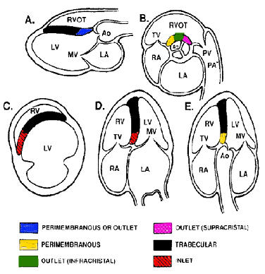
The ventricular septal defect (VSD) is a common congenital heart defect. Due to spontaneous closure during growth the VSD is less often seen in adulthood. VSD’s are divided into three groups: perimembranous, muscular and subarterial. The supracristal outlet VSD is called a subarterial VSD. A subarterial VSD can result in progressive aortic valve insufficiency, due to the lack of the cusp support by the outlet portion of the septum. In some cases even small subarterial VSD’s should be treated surgically. In some circumstances, the septal leaflet of the tricuspid valve can (partially) cover the perimembranous VSD. In these case the images can mimic a septal aneurysm.

VSD

Consequences of VSD

The haemodynamic impact of a VSD-shunt is determined by the size of the VSD, and the pressure differences between right and left cardiac chambers. In a large VSD there will be no flow obstruction between the left and right chamber and the pressures will be equal. This high flow phenomenon often results in pulmonary hypertension. As a result the shunt can become bidirectional or even reversed, this is called Eisenmenger syndrome. A moderately large VSD will not very often lead to resistance pulmonary hypertension. However, in cases of a large shunt (>1:2) it can result in volume load for the left chamber. This leads to dilatation of the left atrium and left ventricle. In small VSD’s, there will be a small shunt without significant volume load of the left chamber. The flow velocity measured over the VSD reflects the pressure gradient. Hence in small VSD the gradients will be high (>4m/s) and with large shunts it will be low or even bidirectional in Eisenmenger.

The information above comes from Echocardiografie.nl. Last changed on: 6 September 2023.