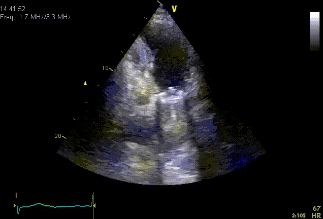
Artifacts are illusions that are projected onto an ultrasound image. It is important to recognize these in order to distinguish between true structures and optical illusions to avoid misdiagnosis. Below is a list of the most common artifacts in alphabetical order.
Aliasing
If the Doppler shift rises above the Nyquist limit. This leads to an opposite blood flow direction seen due to a too low PRF (pulse repetition frequency).
Enhancement
An increase in amplitude of the reflectors which are located behind a weak reflector. For example, in case of pleural or pericardial effusion, the heart will be imaged better because the moisture increases the sound waves.
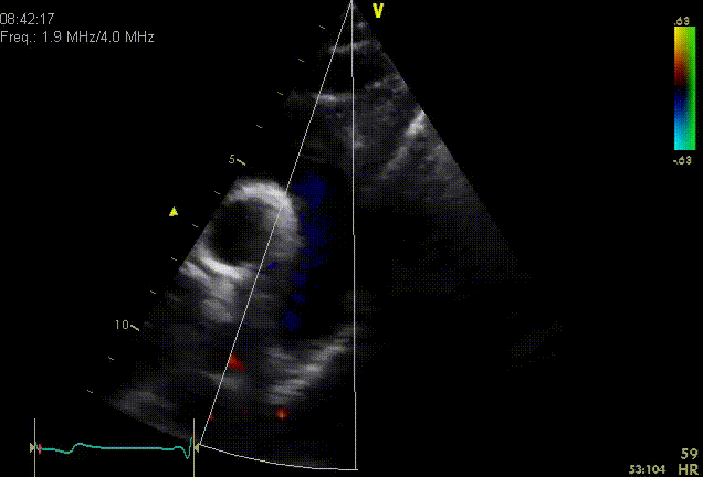
Ghosting
In Color Doppler stains of solid color (red of blue) are painted over regions of the image, as a result of strong of motion of strong reflectors. Transient and not corresponding to expected blood flow.
Mirror artifact
Form of reverberation that a structure is duplicated on the other side of a hard reflector.
Nearfield clutter
Hazy image in the near field as a result of high-amplitude oscillations of the piezo-electric crystals. This could be confused with apical thrombus in the AP4CH view.
Refraction
A change in direction of sound when it strikes a transition boundary of tissue. If the propagation velocity of the first medium is greater than second medium, the resultant angle will be greater than the angle of incidence (think of a pencil in glass of water). Refraction may cause a reflection that is projected laterally from the real object.
Shielding
This is a form of reverberation in which a series of reflecting surfaces are situated very close to each other leading to a form that looks like a comet tail. This phenomenon is especially visible in prosthetic valves.
Partial volume
The sound beam is 3D and the object is imaged as 2D. Tissue that can be imaged is located above / below/behind the object. This is common in circular structures such as pericardium or atria and can be prevented by scanning from another point so you can image the object differently.
Reverberation
Originated by strong reflectors between closely spaced objects. The sound beam is reflected multiple times. Due to the extra distance travelled, the reverberation artefact is depicted deeper.
Shadowing
Decrease in amplitude of the reflectors which are located behind a strong reflector. A strong reflector attenuates the sound distally so that the distal portion is too weak to reflect, and thus results in a dark ?shadow?.
Side lobes
A sound beam has some diverging parts (side lobes). If there is an object outside of the beam, but is affected in a side lobe, this object will be observed as if it was lying centrally in the beam. The echoes from side lobes are therefore not depicted in the correct place.
Speckle
By interference of scattering sound waves in tissue it provides a detailed misleading image resolution (texture within tissue).
Speed error
Axial structures are misplaced as the assumed value of propagation velocity (1540m/s) is incorrect. If the propagation velocity is larger than 1540m/s, the calculated distance to the reflector is too small so that the reflector is shown closer to the transducer. On the other hand, if the propagation speed is smaller than 1540m/s, the reflector will be shown further from the transducer.