Dobu stress echo

Ischemia testing: DSE to detect ischemia

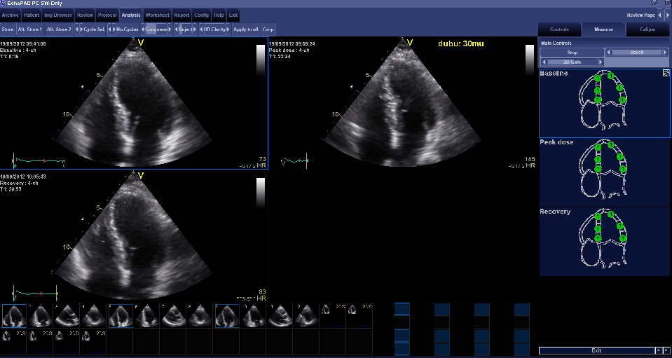
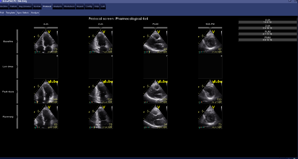
A stress echo is a study which evaluates the function of the LV in rest and during exercise. Ischemia can be provoked by stimulation of the heart by Dobutamine via a drip. Dobutamine is a catecholamine with predominant β-receptor stimulation, which causes positive inotropic, chronotropic and dromotropic effects on the heart. Dobutamine has a half-life of approximately 2 minutes. The inotropic and chronotropic effects can induce myocardial ischemia in case of significant stenosis of a coronary artery. Ischemia leads to hypo- or akinesia in the supplying territory of the artery. Echo has the ability tot detect these new wall motion disorders. Low dose DSE is also used to detect contractile reserve in damaged myocardium after infarction. The test performance for the detection of coronary stenosis is similar to myocardial perfusion imaging and superior to exercise ECG. Initially, images are made in rest: PLAX, PSAX pm, AP4Ch and AP2Ch. During dobutamine infusion, the patient should be monitored using a 12-channel ECG and blood pressure monitor. With each step of increasing dose of dobutamine (every three minutes, see table), an ECG and blood pressure measurement should be performed. A DSE test is safe, but can cause symptoms such as: palpitations, chest pain, dizziness or light-headedness. The dobutamine infusion should be terminated when the target heart rate [(220-age) x 0.85] is reached, serious complications or incidents occur, demonstrable new wall motion abnormalities in more than one segment are observed, or with an increase in end-systolic volume. In cases where the target heart rate has not yet been reached, one should consider to add atropine 0.25mg i.v., also be administered with a maximum of 1 mg (at intervals of 1 minute). At the target heart rate echocardiographic imaging using PLAX, PSax pm, AP4Ch and AP2Ch has to be performed exactly as the rest images to allow for proper comparison. This requires a strict routine of the sonographer. The last step is to ensure that the heart rhythm is back to normal.

DSE 1

DSE 2

DSE at low-gradient aortic stenosis

Low-gradient aortic stenosis is defined as a severe aortic valve stenosis (Effective Orifice Area (EOA) <1.0cm²) with a transvalvular pressure gradient  <30mmHg. A low-gradient aortic stenosis mostly occurs in patients who have a LV dysfunction with a decreased ejection fraction. The calculated EOA can be underestimated in these group of patients because the effect of a low stroke volume on valve opening. A malfunctioning LV can provide insufficient pressure to open the calcified aortic valve. In a fixed or “true stenosis” in which the stroke volume increases, the gradient across the valve increases proportionately. In some patients, the observed increase in stroke volume results in just a limited increase in pressure gradient across the aortic valve. This phenomenon is called a ?pseudo stenosis?. DSE is a tool to distinguish between a true stenosis and a pseudo-stenosis. Patients with pseudo stenosis show an increase in the calculated EOA and a decrease in resistance of the valve in response to an increase in stroke volume. This reaction differs from patients with a true severe aortic stenosis, in whom a dobutamine-induced increase in transvalvular flow results in a proportional increase of the mean transvalvular gradient, resulting in an unchanged EOA. When by increasing stroke volume, an increase in EOA >0.3cm² and a small change in gradient is measured, there appears to be an over-estimation of the severity of the aortic valve stenosis (= pseudo stenosis). A DSE study to evaluate a low-gradient aortic stenosis uses low-dose dobutamine. There study will start with 5µg / kg / min, and gradually increase to up to a maximum dose of 20µg / kg / min. A DSE is also able to provide information on the presence of contractile reserve in patients with a low gradient low EF sever aortic stenosis. Contractile reserve has a predictive value regarding mortality in surgery for aortic valve replacement. Studies have shown that with contractile reserve the perioperative mortality rate calculates 5-8% as compared to 30% in patients without contractile reserve.

Indications and contraindications for DSE

Indications for DSE Contraindications for DSE
suspected coronary artery disease Acute myocardial infarction (≤4-10 days)
Unable to perform exercise testing Unstable angina pectoris
Known coronary artery disease for determining ischemia before and after revascularization Known relevant coronary artery main trunk stenosis
Known coronary artery disease for identifying areas of ischemia Congestive heart failure
Preoperatively, risk estimate, with large myocardial infarction. severe tachyarrhytmia
Demonstration of viability severe valve stenosis
  Hypertrophic obstructive cardiomyopathy
  Acute pericarditis, myocarditis, endocarditis
  aortic dissection

Table of dobutamine concentration

Relation dobutamine concentration on weight and the pump speed
  Time in minutes        
  0 0 3 6 9
  Dobutamine in µg/kg/min (250mg Dobutamine in 50ml)
  5 10 20 30 40
Weight (kg) ml/hour        
44 2.6 5.3 10.5 15.7 20.9
46 2.8 5.6 11.2 16.8 22.4
48 2.9 5.8 11.6 17.4 23.2
50 3.0 6.0 12.0 18.0 24.0
52 3.1 6.2 12.4 18.6 24.8
54 3.2 6.5 13.0 19.4 25.6
56 3.4 6.8 13.6 20.4 27.2
58 3.5 7.0 14.0 21.0 28.0
60 3.6 7.2 14.4 21.6 28.8
62 3.7 7.4 14.8 22.2 29.6
64 3.8 7.6 15.2 22.8 30.4
66 4.0 8.0 16.0 24.0 32.0
68 4.1 8.2 16.4 24.6 32.8
70 4.2 8.4 16.8 25.2 33.6
72 4.3 8.6 17.2 25.8 34.4
74 4.4 8.8 17.6 26.4 35.2
76 4.6 9.2 18.4 27.6 36.8
78 4.7 9.4 18.8 28.2 37.6
80 4.8 9.6 19.2 28.8 38.4
82 4.9 9.8 19.6 29.4 39.2
84 5.0 10.0 20.0 30.0 40.0
86 5.1 10.2 20.4 30.6 40.8
88 5.3 10.6 21.2 31.8 42.4
90 5.4 10.8 21.6 32.4 43.2
92 5.5 11.0 22.0 33.0 44.0
94 5.6 11.2 22.4 33.6 44.8
96 5.8 11.6 23.2 34.8 46.4
98 5.9 11.8 23.6 35.4 47.2
100 6.0 12.0 24.0 36.0 48.0
102 6.1 12.2 24.4 36.6 48.4
104 6.2 12.4 24.8 37.2 49.6
106 6.4 12.8 25.6 38.4 51.2
108 6.5 13.0 26.0 39.0 52.0
110 6.6 13.2 26.4 39.6 52.8

Click for PDF version of table of dobutamine concentration

The information above comes from Echocardiografie.nl. Last changed on: 4 September 2023.