Aorta

The aortic annulus should be measured at midsystole from inner edge to inner edge. All other aortic root measurements (i.e., maximal diameter of the sinuses of Valsalva, the sinotubular junction, and the proximal ascending aorta) should be made at end-diastole, in a strictly perpendicular plane to that of the long axis of the aorta using the leading edge-to-leading edge (L-L) convention.

Aortic root dimensions in normal adults

Aortic measures according to sex
  ALL MEN WOMEN
Aortic annular diameter, mm 20.4 ± 2.3 21.2 ± 2.2 19.5 ± 2.1*
BSA-indexed annular diameter, mm/m² 11.6 ± 1.4 11.3 ± 1.3 11.9 ± 1.4
Height-indexed annular diameter, mm/m 12.2 ± 1.2 12.3 ± 1.2 12.2 ± 1.3
Aortic SoV diameter, mm 30.8 ± 3.9 32.2 ± 3.7 29.3 ± 3.6*
BSA-indexed aortic SoV diameter, mm/m² 17.6 ± 2.6 17.2 ± 2.5 18.0 ± 2.6*
Height-indexed aortic SoV diameter, mm/m 18.5 ± 2.3 18.6 ± 2.3 18.4 ± 2.3*
Aortic STJ diameter, mm 26.6 ± 3.7 27.7 ± 3.7 25.5 ± 3.3*
BSA-indexed aortic STJ diameter, mm/m² 15.2 ± 2.4 14.8 ± 2.3 15.6 ± 2.4*
Height-indexed aortic STJ diameter, mm/m 16.0 ± 2.2 16.0 ± 2.2 15.9 ± 2.2
Prox. ascending ao (cm/m²)   ≤ 1,7 ≤ 1,9

Data are expressed as mean 6 SD or as number (percentage)
*P < .05, men versus women.
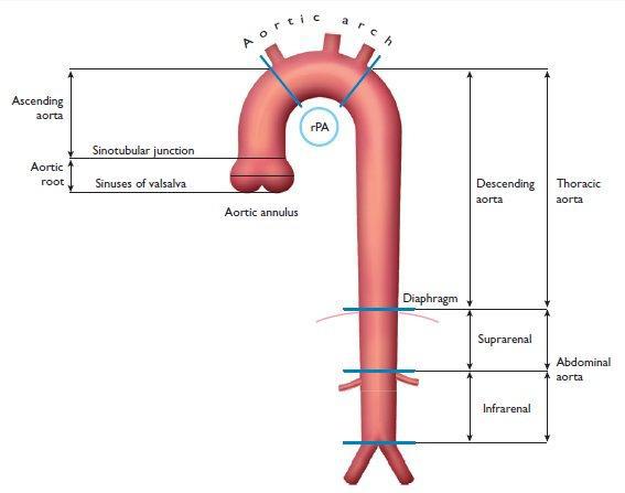
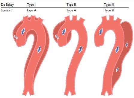
Anatomy of aorta
Anatomy of aorta

Ascending aorta dilation

Ascending aorta dilation


Evaluation aortic dissection

Checklist
Location of dissection
Type A (involvement of ascending aorta)
Type B (no involvement of ascending aorta)
Location of intimal tear
Location of re-entry and possibly another tear
Involvement of aortic arch and outgoing vessels
Differentiation between true and false lumen
Involvement of coronary arteries
Presence of aortic valve insufficiency
Pericardial effusion
LV function

Classification of aortic dissection


Classification of aortic dissection


Differentiation between true and false lumen

Differentiation
In M-mode moves the flap to the false lumen in systole.
Spontaneous echo contrast and thrombus can be seen in the false lumen.
On color Doppler, flow is shown slower systolic by secondary or re-entry to tear the false lumen.
The false lumen (especially in chronic dissections) tends to be larger in comparison to the true lumen.

Evaluation of coarctation aortae

Parameter Instrument commentary
Location Color doppler With the origin of the carotid and subclavian artery are landmarks for locating the coarctation.
Velocity profile Continuous wave Remember that collateral will reduce the systolic speed but the diastolic gradient persists. In the presence of diastolic forward flow is spoken of a hemodynamically significant coarctation.
    Typically CW Doppler signal of descending aorta with diastolic forward flow fitting in hemodynamically significant coarctation

The information above comes from Echocardiografie.nl. Last changed on: 11 September 2023.